⚠️ Updated Lecture Available! This version of the Mitral Stenosis lecture is from an earlier year. For the new, improved, and fully visual 2025 edition, watch it here: 👉 NEW 2025 Mitral Stenosis Lecture: This original video is still available for reference, but the updated lecture includes: New 2025 echo visuals Updated explanations Clearer Doppler + 2D imaging examples Registry-focused breakdowns Better flow + improved teaching clarity If you're studying for boards or clinicals, I strongly recommend watching the new version. In this cardiac ultrasound tutorial, we break down mitral valve stenosis from an echocardiography perspective. Learn how to identify the key 2D, M-mode, and Doppler findings that confirm mitral stenosis, Whether you’re a cardiac sonography student, RDCS preparing for boards, or an ultrasound professional looking to refine your echo skills, this video provides a clear, step-by-step explanation of what mitral stenosis looks like on echo and how to properly evaluate its severity. Chapters: 📌 Cardiac Ultrasound Topics Covered: • What mitral valve stenosis is and how it affects cardiac function • Classic 2D echo findings (hockey-stick anterior leaflet, thickened leaflets, reduced mobility) • Doppler evaluation: pressure half-time, mean gradients, and diastolic flow patterns • Techniques for optimizing apical and parasternal views • How to differentiate mild, moderate, and severe stenosis on echo • Common pitfalls and how to avoid mis-measurements • Clinical significance and correlation with symptoms 🎯 Perfect for: • Cardiac sonographers • RDCS / CCI exam prep • Echocardiography students • Healthcare professionals learning cardiac imaging 👍 Like, subscribe, and share for more cardiac ultrasound education, echo tips, and pathology breakdowns! #ValvularHeartDisease #Echocardiography #EchoEducation #EchoNotebook #EchoLearning #EchoStudent #EchoTraining #CardiacSonography #SonographyEducation #CardiacUltrasound
- 236Просмотров
- 2 недели назадОпубликованоThe Echo Classroom: Minks Koroma RDCS, RDMS, RVT
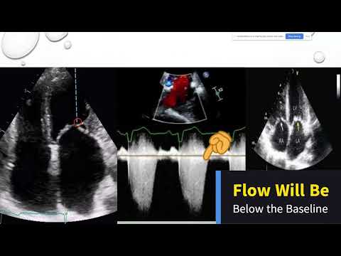
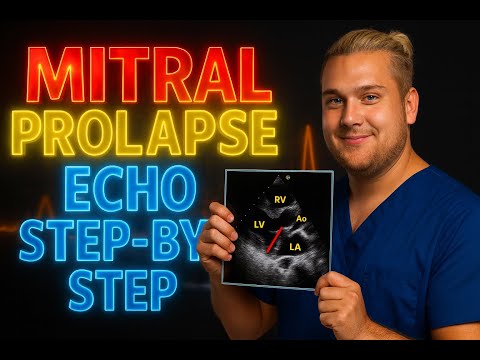
8.2 How to Assess Mitral Stenosis on Echocardiography | Step-by-Step Guide for Echo Students
Похожее видео


Популярное
Новини